QIBEBT Developed Multifunctional Virus-mimetic Nanostructure by Self-assembly
Molecular self-assembly is ubiquitous in nature and fabricates a variety of biological structures. Viruses represent a kind of highly assembled symmetrical architectures and are ideal nanoscale platform to construct the bio-mimetic nanocomposites, which can be found wide applications in biosensing, gene delivery, novel nanometer devices, biomedical imging and tumor therapy.
By mimicking the filamentous phage, graduate student, Miss WANG Fei from the Laboratory for Biosensing, Qingdao Institute of Bioenergy & Bioprocess Technology (QIBEBT) and her coworkers successfully constructed multifunctional virus-mimetic nanostructure using layer-by-layer self-assembly strategy, realizing the simultaneous colorectal tumor cell SW620 specifically fluorescent imaging and photothermal therapy (PTT). This work was published online in Scientific Reports.
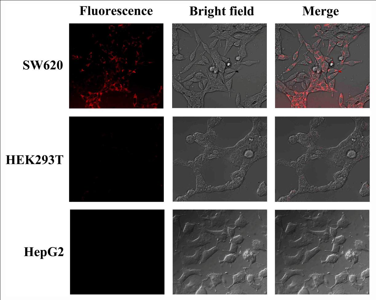
The researchers first selected the SW620 cell-specific peptide from the f8/8 landscape phage library. The dye molecules and isolated specific pVIII fusion coat proteins with dipole property were sequentially self-assembled on the surface of the Au@Ag heterogeneous nanorods, which exhibited typical near IR spectrum at 803 nm. For the as-prepared bio-mimetic nanostructure, the specific octapeptide of pVIII fusion proteins arranged outwardly (Fig. 1). The resultant nanocomposites had excellent biocompatibility and showed cancer cell-specific fluorescence detection (Fig. 2). Moreover, the nanocomposites were capable of specifically ablating cancer cells in the light power of 4 W/cm2 (Fig. 3), which is lower than 10 W/cm2 reported for gold nanorods and silver nanoparticles.
This research was led by Prof. LIU Aihua from the Laboratory for Biosensing, QIBEBT. This work was supported by National Natural Science Foundation of China.
 |
|
Fig. 1. Schematic preparation of the virus-mimetic nanostructure by layer-by-layer self-assembly. |
 |
|
Fig. 2. Confocal fluorescence images of target cells SW620 and control cells (HEK293T and HepG2) after incubation with the virus-mimetic nanostructures. |
 |
|
Fig. 3. The cell viability of target and control cells was tested under irradiation for different time with an 808 nm laser after incubation with the virus-mimetic nanostructures. (Images by Laboratory for Biosensing, QIBEBT) |
Reference:
F. Wang, et al., Scientific Reports 2014, 4, 6080. http://www.nature.com/srep/2014/141028/srep06808/pdf/srep06808.pdf
Contact:
Prof Dr. LIU Aihua
Email: liuah (AT) qibebt.ac.cn